
Formulation and Delivery - Biomolecular
Category: Late Breaking Poster Abstract
 photo")
Yingchang Ma, PhD (she/her/hers)
PhD student
University College London
London, England, United Kingdom
Yingchang Ma, PhD (she/her/hers)
PhD student
University College London
London, England, United Kingdom
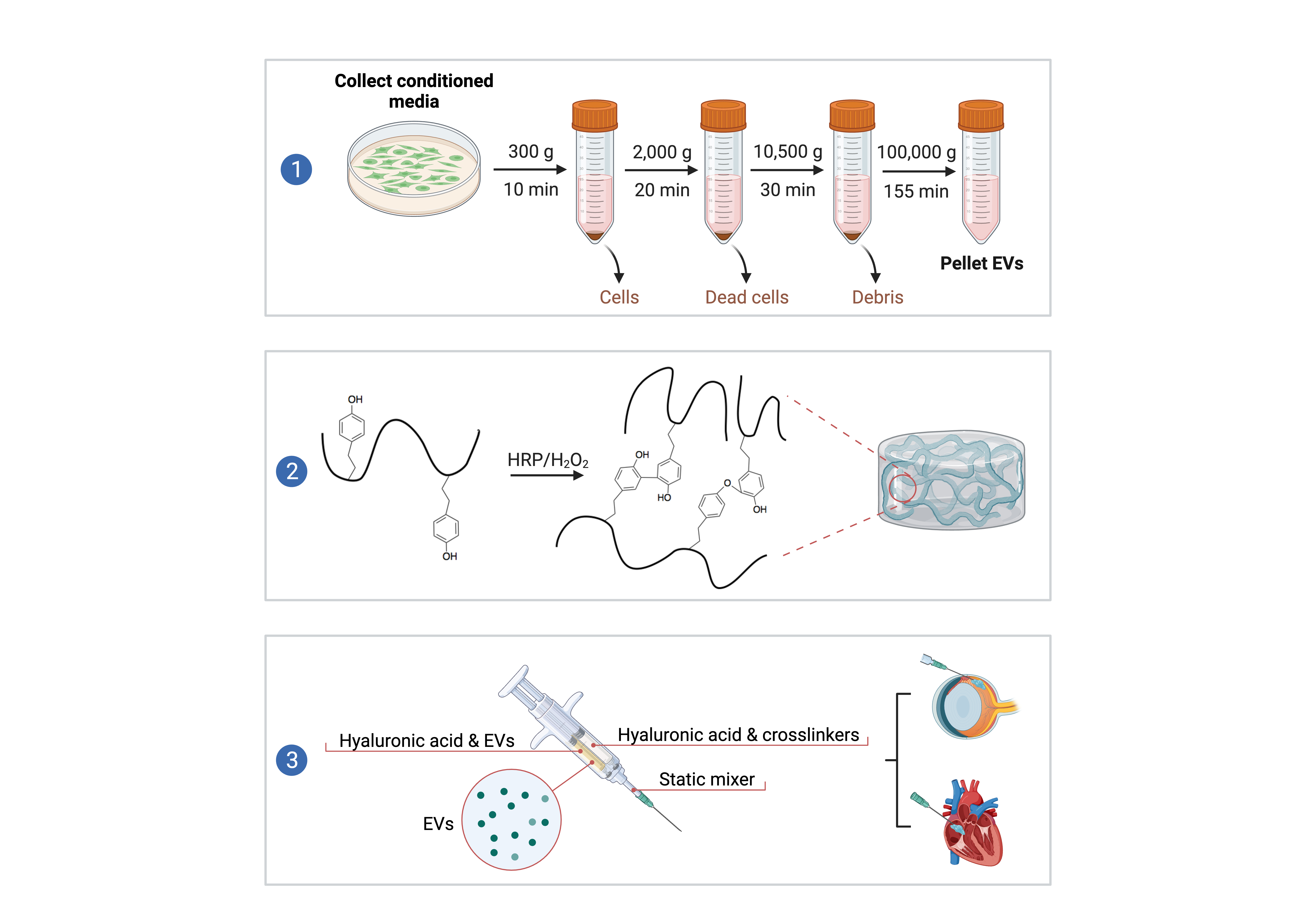 Figure 1. 1) EV isolation by ultracentrifugation; 2) Synthesis of HA-Tyr conjugates; 3. Preparing EV-loaded hydrogel in double syringe.
 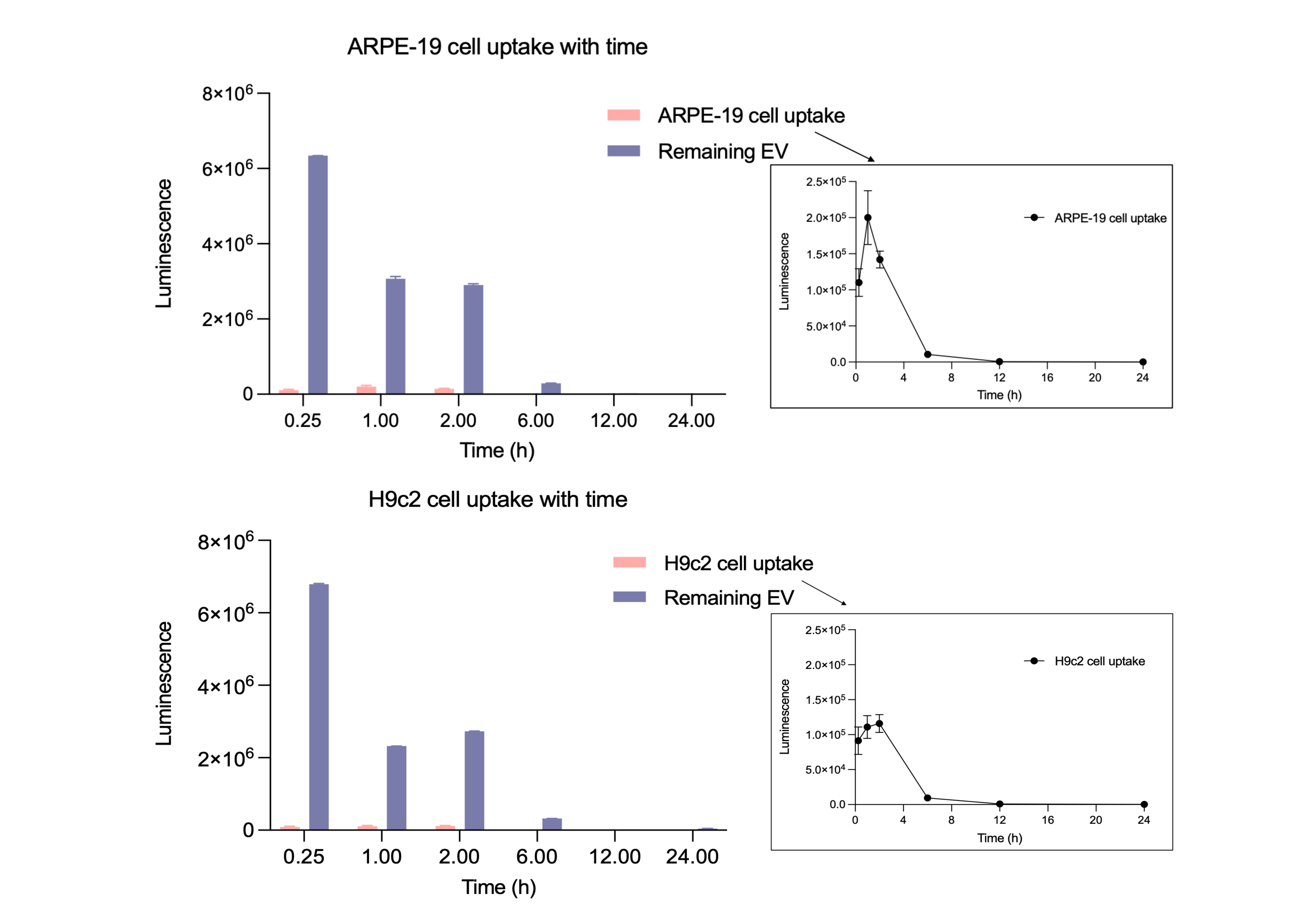 Figure 2. Kinetic study. ARPE-19 and H9c2 cells were incubated with EVs (1E11 particles/mL) for different incubation times (15 min, 1, 2, 6, 12, and 24 h) at 37 °C. EV uptake was then quantified by luciferase assay. Kinetics showed that luciferase activity in acceptor cells increased over time, with a 1% spontaneous rate at 1 h.
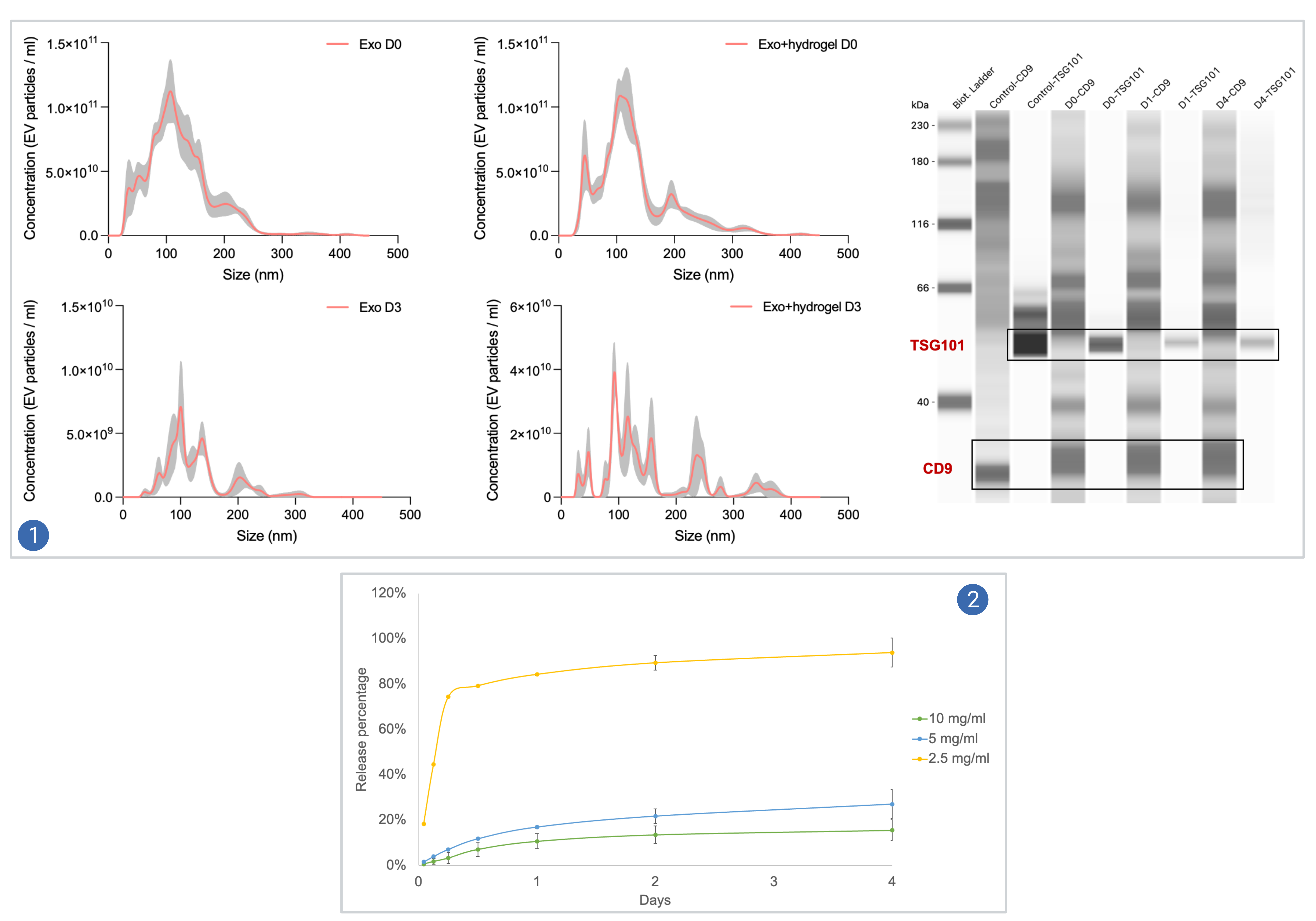 Figure 3. 1) The stability of EVs loaded in the hydrogels was determined by comparing the particle concentrations and protein expressions with non-loaded EVs. The particle concentration was analysed by NTA (left), and the expression of EV markers (CD9, TSG101) was analysed by simple western blot (right); 2) The cumulative release profiles of EVs from hydrogels. Short-term release (1, 3, 6, 12 h) was determined by NTA, and long-term release (1, 2, 4 days) was quantified by CD9-ELISA kit.