
Formulation and Delivery - Biomolecular
Category: Poster Abstract
 photo")
Ketki Velankar, MS (she/her/hers)
Student
Duquesne University - Graduate School of Pharmaceutical Sciences
Pittsburgh, Pennsylvania, United States
Ketki Velankar, MS (she/her/hers)
Student
Duquesne University - Graduate School of Pharmaceutical Sciences
Pittsburgh, Pennsylvania, United States
Wen Liu, Ph.D.
Allegheny Health Network Cancer Institute
Pittsburgh, Pennsylvania, United States
Paul Hartmeier, BS (he/him/his)
Graduate Student
Duquesne University
Pittsburgh, Pennsylvania, United States
Benjamin Clegg, BS
Duquesne University
Pittsburgh, Pennsylvania, United States
Ellan Gawalt, Ph.D.
Duquesne University
Pittsburgh, Pennsylvania, United States
Yong Fan, Ph.D.
Allegheny Health Network Cancer Institute
Pittsburgh, Pennsylvania, United States
Wilson Meng, Ph.D. (he/him/his)
Duquesne University
Pittsburgh, Pennsylvania, United States
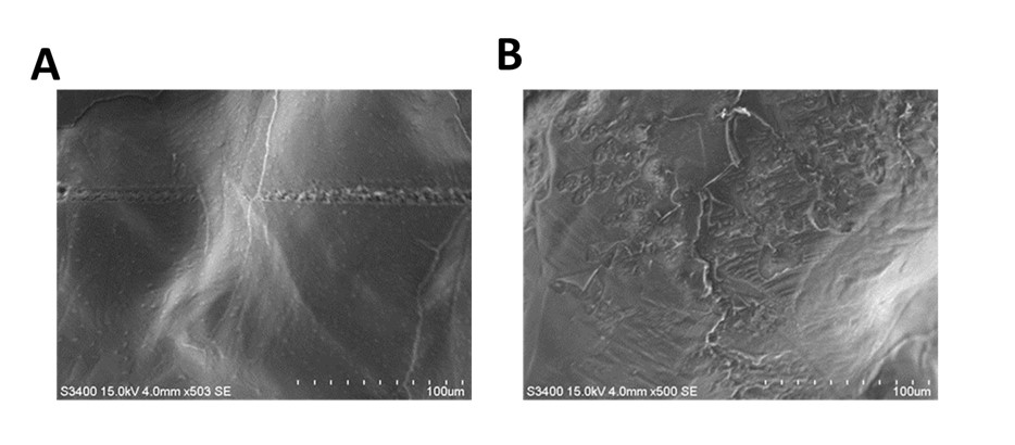 Fig.1 Scanning electron microscopy (SEM) imaging of EAKbt-avidin formulation. SEM images of (A) EAKbt and (B) EAKbt+avidin showing denser peptidic assembly in the latter.
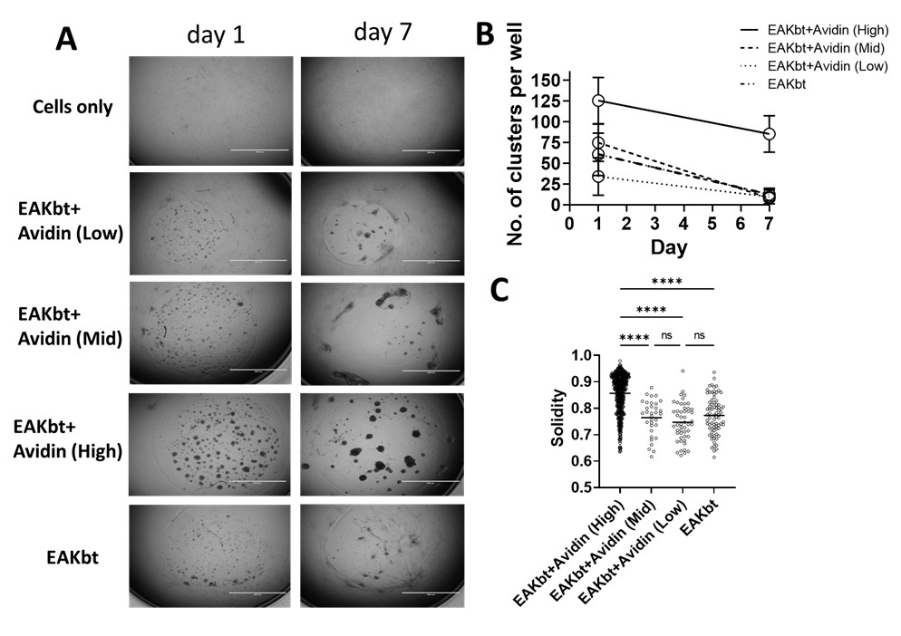 Fig. 2 Formation and characterization of reticular clusters of human lymphatic fibroblasts. (A) Representative brightfield microscopic images showing cell cluster formation in the presence of EAKbt-avidin as a function of avidin concentration; (B) Effect of EAKbt+Avidin on retaining clusters cultured till day 7; (C) Image analysis of clusters (day 7) showing higher solidity in high avidin group representing uniform outer morphology (absence of adhering (2D), elongated cells) indicative of 3D organization; **** represents p < 0.0001 (One-way ANOVA with multiple comparisons).
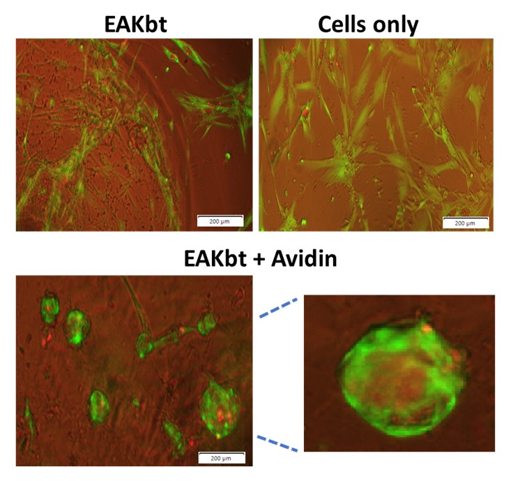 Fig. 3 Live-dead staining of cell clusters. Fluorescent microscopy images of live-dead staining for cells depicting live (green) and dead (red) in the biomaterial formulation.