
Formulation and Delivery - Biomolecular
Category: Late Breaking Poster Abstract
 photo")
Hend Mohamed Abdel-Bar, PhD (she/her/hers)
Associate Professor of Pharmaceutics
University of Sadat City
Sadat City, Al Minufiyah, Egypt
Hend Mohamed Abdel-Bar, PhD (she/her/hers)
Associate Professor of Pharmaceutics
University of Sadat City
Sadat City, Al Minufiyah, Egypt
Isra H. Ali, Ph.D. (she/her/hers)
University of Sadat City
Sadat City, Al Minufiyah, Egypt
Mohamed Hamdi, Ph.D. (he/him/his)
University of Sadat City
Sadat City, Al Minufiyah, Egypt
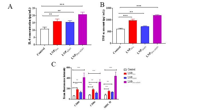 Figure 1. The co-delivery of doxorubicin and nZnO into lipid nanoparticles induced macrophage polarization. After 24h of J774 cells seeding in 24-well plate at density of 50K/ well, cells were treated with either LNPDox, LNPnZnO or LNPDox-nZnO for 24 h. Following the supernatant collection and centrifugation, the concentrations of IL-6 (A) and TNF-α (B) were measured using ELISA. Cells harvested by trypsin-EDTA were centrifuged at 1750 rpm for 3 min at 4 °C then stained with either anti-mouse CD80-FITC monoclonal antibody (1: 200 v/v), anti-mouse CD86-FITC monoclonal antibody (1: 200 v/v) or anti-mouse MHC II-FITC monoclonal antibody (1: 200 v/v) for 30 min at 4 °C. The expression of CD80, CD86 and MHC II was quantified using the fluorescence in FL1. The expression of each costimulatory marker was presented as MFI marking also higher marker values with LNPDox-nZnO (C). Data points represented mean and SD (n=3). Statistical analysis was performed using One-way ANOVA followed by Tukey’s post-test **p < 0.01, ***p < 0.001.
 Figure 2. The co-delivery of doxorubicin and nZnO into lipid nanoparticles induced ATP, HMGB1 secretion, surface calreticulin expression and consequently macrophage phagocytosis. The effect of the proposed system on ATP and HMGB1 secretion. 4T1 cells were seeded in 24-well plate at density of 50K/ well for 24 h, then treated with LNPDox, LNPnZnO or LNPDox-nZnO for 24 h. Afterwards, the supernatant was collected and the cellular debris removed by centrifugation. The concentration of acellular ATP was measured by luciferase based ATP detection reagent (A) and HMGB1 release in the culture media was quantified using ELISA (B). Cells were collected by trypsin-EDTA, centrifuged at 1750 rpm for 3 min then stained using rabbit anti-human calreticulin antibody (1:50 v/v) followed by donkey anti-rabbit IgG-FITC secondary antibody (1:200). The fluorescence signals were measured in FL1 channel. Gates were plotted based on the isotype. Surface calreticulin expression was expressed as mean fluorescence intensity (MFI) (C). The treated 4T1 cells were collected and co cultured with J774 macrophage cells for 6 h. Cells were harvested and stained with anti-mouse CD45 monoclonal before being acquired on a FACs Calibur flow cytometer. Relative MFI of CD45+ J774 cells is shown in (D). LNPDox, LNPnZnO or LNPDox-nZnO elevates ATP concentrations, calreticulin and macrophage phagocytosis over untreated cells. However, only doxorubicin loaded formulae were able to induce HMGB1 release. Data points represent mean and SD (n=3). Statistical analysis was performed using One-way ANOVA followed by Tukey’s post-test *p < 0.05, **p < 0.01, ***p < 0.001, ns nonsignificant.