
Discovery and Basic Research
Category: Late Breaking Poster Abstract
Yong-Bok Lee
Chonnam National University
Gwangju, Kwangju-jikhalsi, Republic of Korea
Ji-Hun Jang
Chonnam National University
Gwangju, Kwangju-jikhalsi, Republic of Korea
Seung-Hyun Jeong
Sunchon National University
Suncheon-si, Cholla-namdo, Republic of Korea
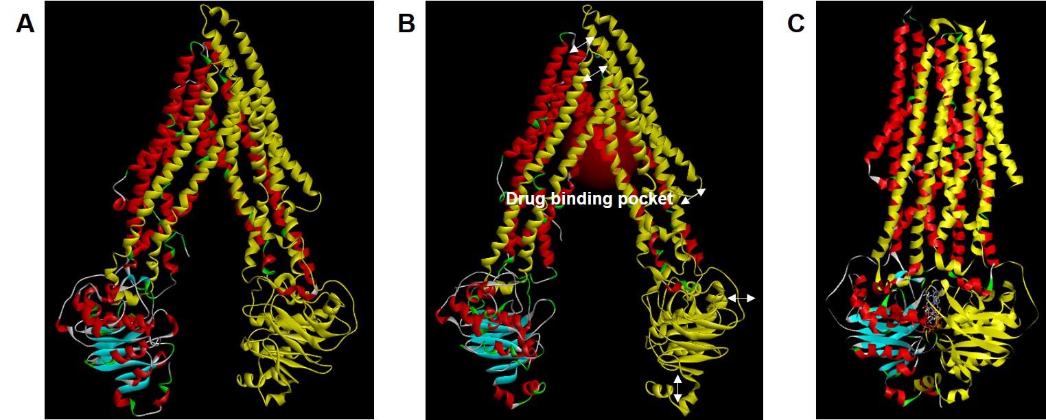 Figure 1. Molecular structures of P-glycoprotein (P-gp) in apo-state (A), ligand-docked state (B), and outward-facing state (C). Arrows indicate major conformational sites in the ligand-docked state compared to the apo-state. For easy comparison of steric changes in each conformational state of P-gp, cassette 1 is labeled in yellow. In the ligand-docked state, red-labeled region represents drug binding pocket.
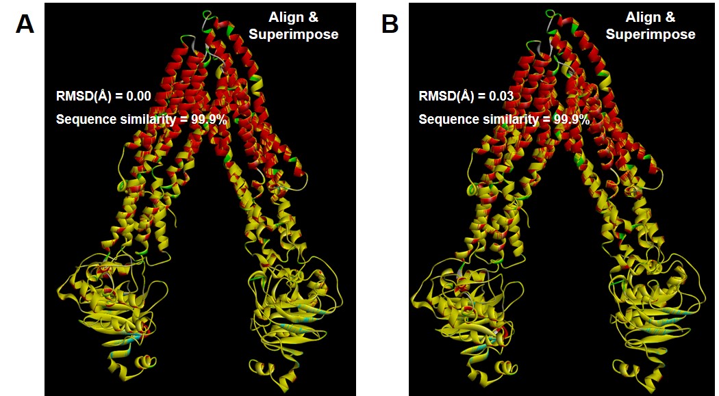 Figure 2. Comparison (alignment and superimpose with wild-type structures) of point polymorphism structures of Ala893Ser (A; ABCB1 2677G>T) and Ala893Thr (B ; ABCB1 2677G>A) in the ligand-docked state of P-glycoprotein. Point polymorphism structures by Ala893Ser/Thr are labeled in yellow.
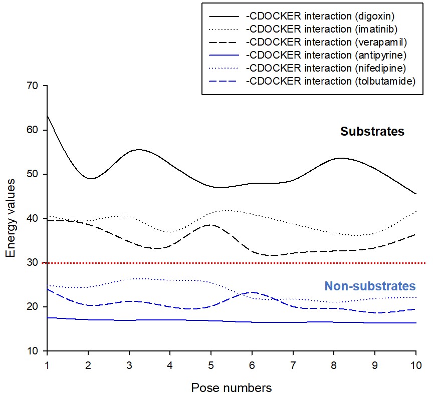 Figure 3. Comparison graph of –CDOCKER interaction energy values according to docking simulation with P-glycoprotein (P-gp) ligand-docked state of drugs corresponding to substrate (digoxin, imatinib, and verapamil) or non-substrate (antipyrine, nifedipine, and tolbutamide) of P-gp. The red dotted line in the graph represents the energy value (30) that approximates the degree of effective substrate interaction with P-gp.