
Formulation and Delivery - Chemical
Category: Late Breaking Poster Abstract

Hongyi Chen
PhD candidate
Fudan University
Shanghai, Shanghai, China (People's Republic)
Hongyi Chen
PhD candidate
Fudan University
Shanghai, Shanghai, China (People's Republic)
Tao Sun, Ph.D.
Fudan University
Shanghai, Shanghai, China (People's Republic)
Chen Jiang, Ph.D.
Fudan University
Shanghai, Shanghai, China (People's Republic)
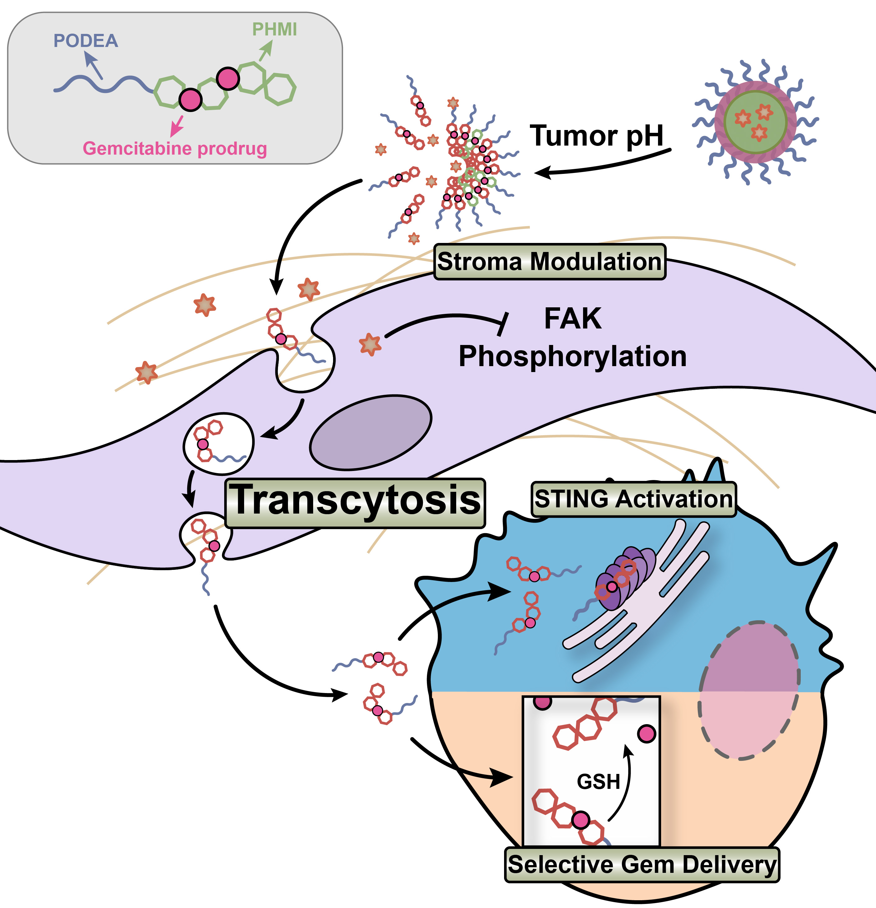 Figure 1. Schematic illustration of the transcytosis mediated tumor penetration, microenvironment acidity and intracellular GSH dual-responsive drug release profile of nanoparticles with the stroma modulation and combined chemo/immunotherapy promotion capacity.
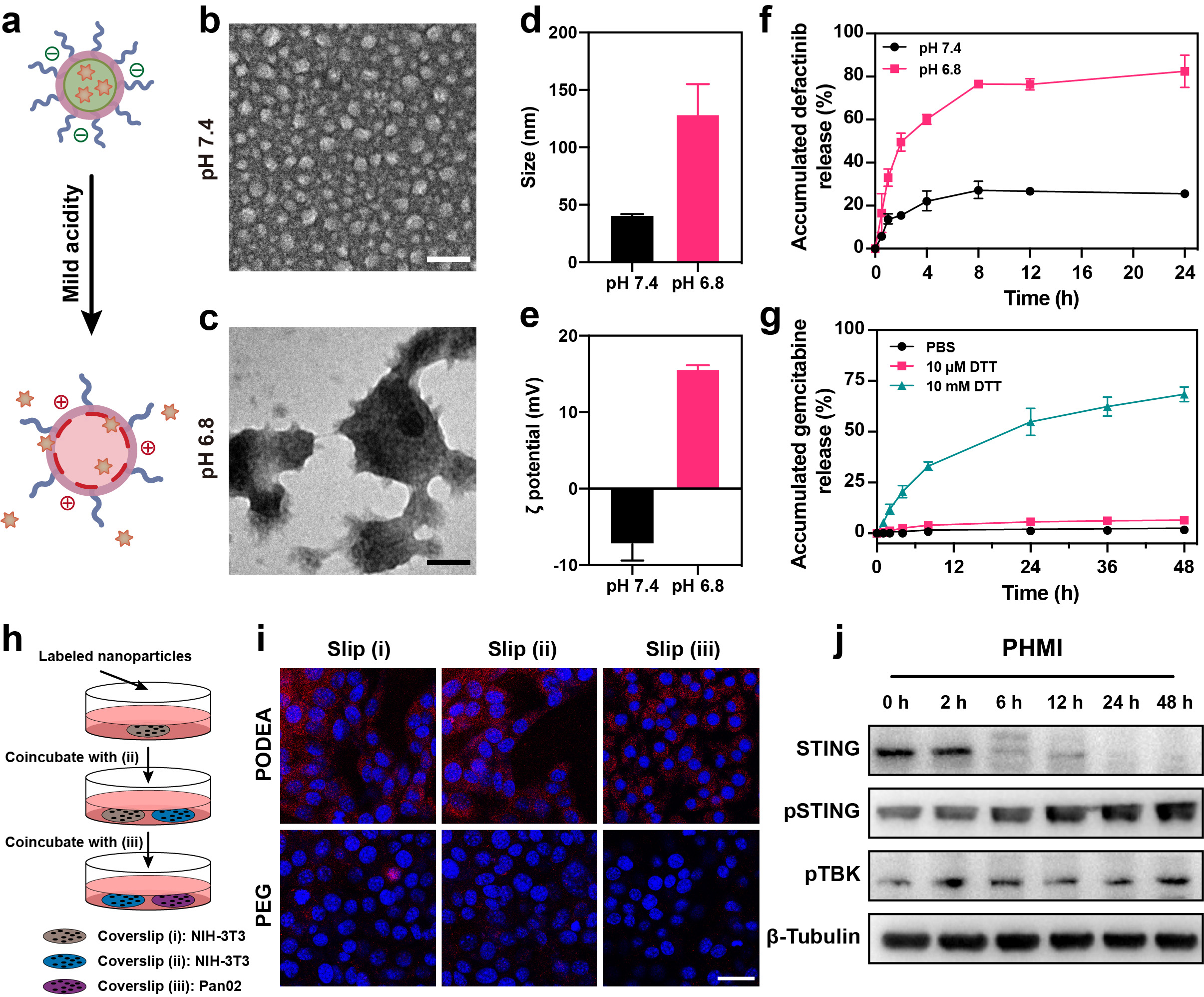 Figure 2. The profiles of nanoparticles in vitro. a) Illustration of mild acidity triggered drug release and charge reversal of the PODEA-Gem-HMI nanoparticles. The TEM images of the nanoparticles at b) pH 7.4 and c) pH 6.8. Scale bars = 100 nm. The change of the d) size and e) ζ potential of the nanoparticles at pH 7.4 and 6.8. f) Release profile of defactinib in PBS 7.4 and 6.8 at 37 °C. g) The release profile of gemcitabine triggered by different concentrations of dithiothreitol in PBS 7.4 at 37 °C. h) Illustration showing the protocol of visualizing transcytosis with coverslip coincubation. i) CLSM images of the cells on different coverslips. Scale bar = 30 µm. j) Western blot of STING, pSTING, and pTBK expression in primary macrophages at different time points after being treated with PODEA-HMI. β-Tubulin was used as the loading control. The data are represented as the means ± SD (n = 3).
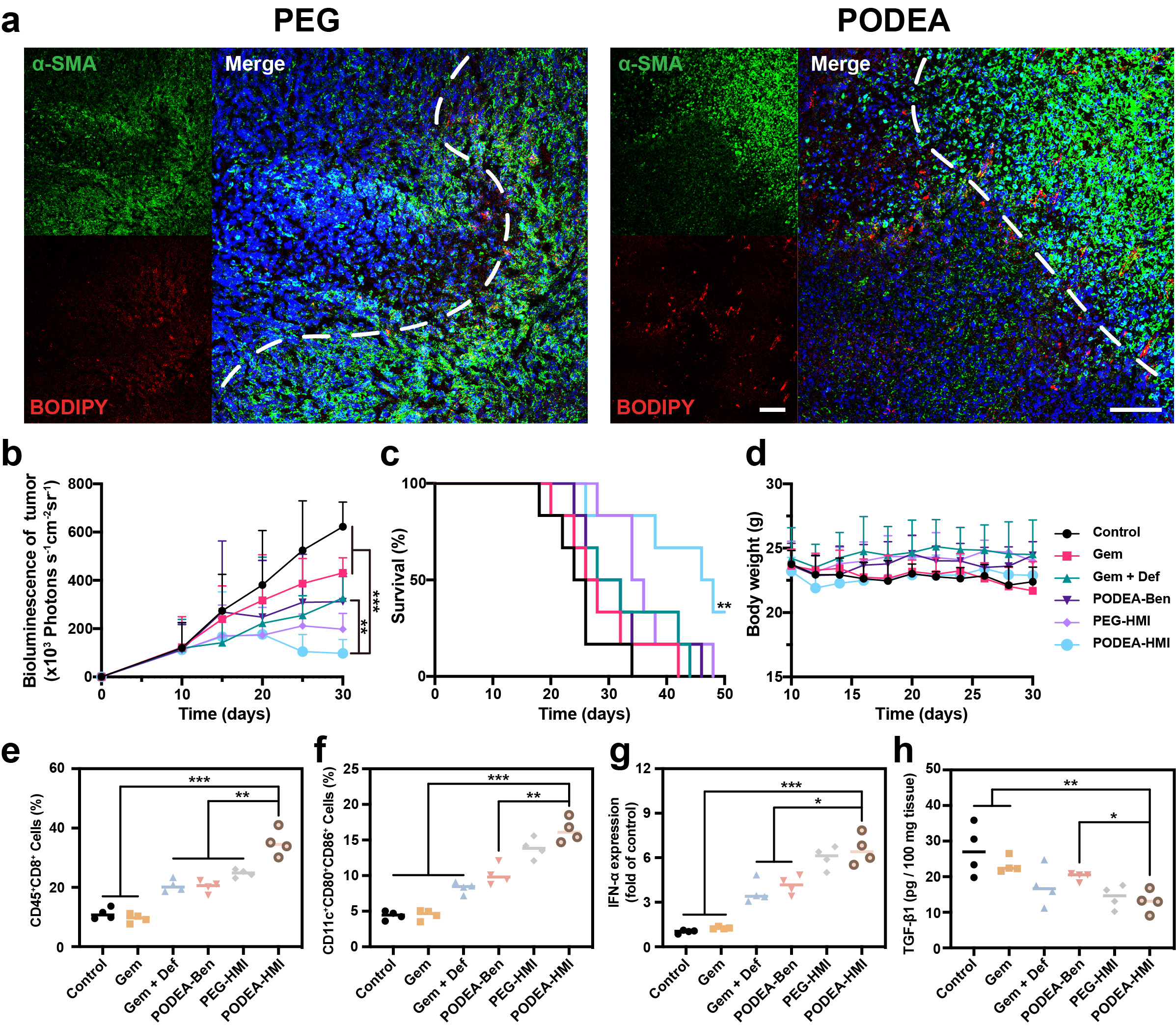 Figure 3. The profiles of nanoparticles in vivo. a) Representative fluorescent images of frozen tumor slices from PDAC-bearing C57 mice. Blue: DAPI for staining cell nucleus; green: Alexa Fluor 488 labeled α-SMA for staining tumor stroma; red: BODIPY 630/650 labeled nanoparticles. Scale bars = 100 µm. b) Tumor volume changes measured by the bioluminescence signal. c) Survival curves of different groups of treatment. d) Body weight recorded 20 days after drug administration. Statistical results of the FACS analysis of e) cytotoxic T cells (CD45+CD4−CD8+) and f) matured DCs (CD11c+CD80+CD86+). g) The expression of IFN-α in the tumor tissue detected by RT-qPCR. h) Expression of TGF-β1 in tumors. The data are represented as means ± SD (n = 4). *, **, and *** denote p < 0.01, p < 0.005, and p < 0.001.