
Formulation and Delivery - Biomolecular
Category: Late Breaking Poster Abstract
 photo")
Esraa Mohamed, PhD (she/her/hers)
PhD Graduate Student
University of Iowa
Iowa City, Iowa, United States
Noah Laird (he/him/his)
University of Iowa
Iowa city, Iowa, United States
Pornpoj Phruttiwanichakun (they/them/theirs)
University of Iowa
Iowa City, Iowa, United States
Douglas Fredericks (they/them/theirs)
University of Iowa
Coralville, Iowa, United States
John M. Femino (they/them/theirs)
University of Iowa
Iowa City, Iowa, United States
Aliasger Salem, Ph.D.
University of Iowa
Iowa City, Iowa, United States
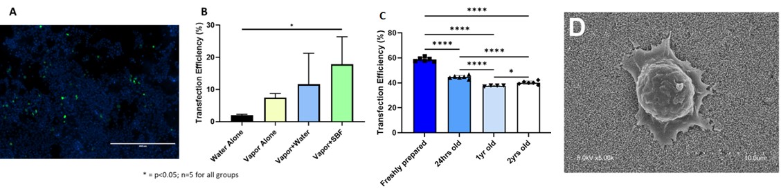 Figure 1: Fluorescence micrograph (A) Transfection efficiency of HEK 293T cells seeded onto gene-activated CPC discs as determined by flow cytometry; Bars represent mean + SD, n=5 for all groups for different treatment methods (B) transfection efficiency of nanoplexes confirming the long term stability of nanoplexes to deliver the pDNA (C) and Micrographs of HEK 293T cells incubated for 4 hours on printed scaffolds mineralized with vapor then simulated body fluid (D).
 .jpg) Figure 2: SEM images of (A) Blank MPs and (B) Insulin loaded MPs and (C) Cumulative release of calcitriol (vitamin D) from CPC scaffolds (Values represent mean + SD, n=6 for all samples).
Figure 2: SEM images of (A) Blank MPs and (B) Insulin loaded MPs and (C) Cumulative release of calcitriol (vitamin D) from CPC scaffolds (Values represent mean + SD, n=6 for all samples)..jpg) Figure 3: (A) CPC scaffold next to collagen ribbon that contains insulin-loaded microparticles, (B) CPC scaffold wrapped in collagen ribbon, (C) CPC scaffold/collagen ribbon during implantation in the defect site (D) X-ray of CPC scaffold/collagen ribbon after implantation.
Figure 3: (A) CPC scaffold next to collagen ribbon that contains insulin-loaded microparticles, (B) CPC scaffold wrapped in collagen ribbon, (C) CPC scaffold/collagen ribbon during implantation in the defect site (D) X-ray of CPC scaffold/collagen ribbon after implantation.