
Discovery and Basic Research
Category: Late Breaking Poster Abstract
Thanh C. Dinh, B.S. (he/him/his)
Florida A&M University
Tallahassee, Florida, United States
Thanh C. Dinh, B.S. (he/him/his)
Florida A&M University
Tallahassee, Florida, United States
Shallu Kutlehria, Ph.D. (she/her/hers)
Florida A&M University
Tallahassee, Florida, United States
Arvind Bagde, Ph.D. (he/him/his)
Florida A&M University
Tallahassee, Florida, United States
Nilkumar (Neal) Patel, Ph.D.
Florida A&M University
Tallahassee, Florida, United States
Aragaw Gebeyehu, Ph.D. (he/him/his)
Florida A&M University
Tallahassee, Florida, United States
Mandip Singh, Ph.D.
Florida A&M University
Tallahassee, Florida, United States
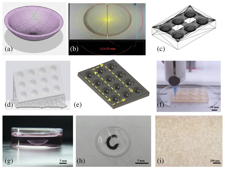 The development of human corneal stroma and base support scaffolds apparatus. (a) Average corneal stroma dimensions modeled in Autodesk Fusion 360. (b) Bright-field image of single well of support scaffold showing its radius. (c) 3D model of 6-well corneal scaffold. (d) Support and raft preparation with Preform software for SLA printing of 12-well scaffold. (e) Printing path of 12-well corneal scaffold. (f) High-throughput printing of 12 corneas with a BIOX printer. (g) Printed cornea after crosslinking and placing within media. (h) Picture of cornea showing its transparency through the letter “C.” (i) Bright-field image of 3D printed cornea using inverted microscope on Day 1
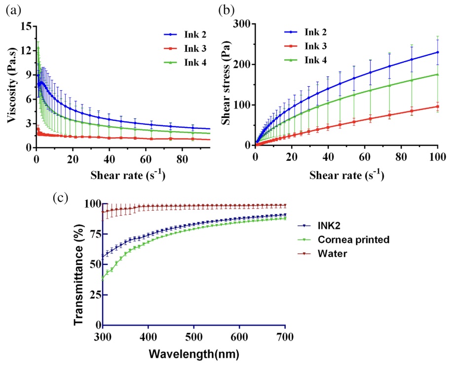 Characterization of bioinks (a) Flow sweep test using rheometer to determine the viscosity at varying shear stress. Ink 2 exhibited higher viscosity as compared to others. (b) Shear stress vs shear rate behavior of bioinks showing shear thinning. (c) Light transmittance of bio-ink 2, 3D printed cornea at varying wavelengths compared with control (water).
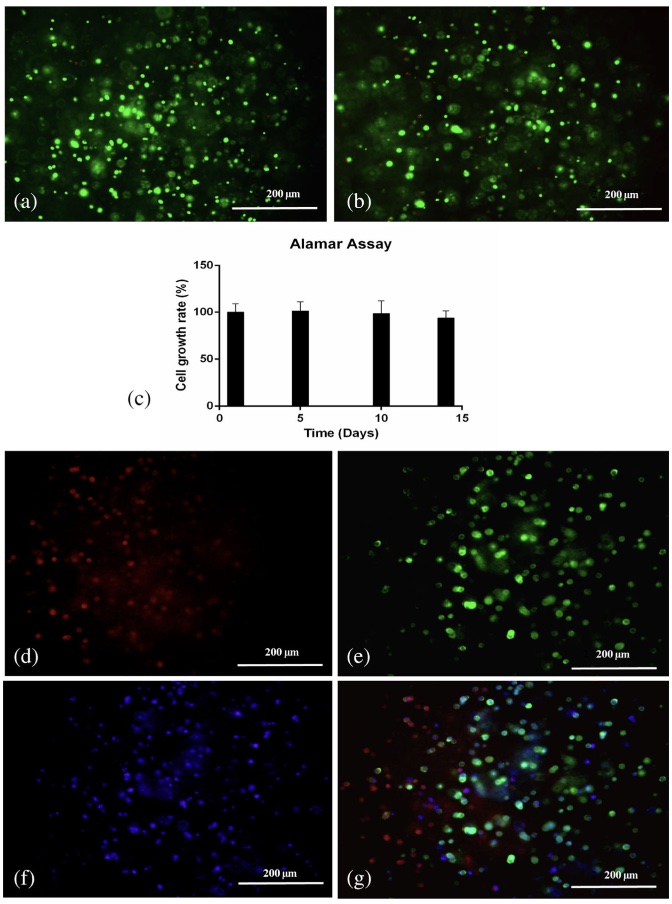 Evaluation of cell viability and immunofluorescence staining of 3D bioprinted corneas. (a) Live dead assay in 3D printed corneas bearing human corneal keratocyte (HCK) cells on Day 1 (merged image). (b) Live dead assay in 3D printed corneas bearing HCK cells on Day 14 (merged image). (c) Alamar assay on 3D printed corneas on Days 1, 5, 10, and 14. Immunofluorescence staining of 3D printed corneas using a fluorescent microscope for (d) fibronectin (red), (e) F-actin (green), (f) DAPI (blue), and (g) merge image