
Bioanalytics - Chemical
Category: Late Breaking Poster Abstract

Shuhua Bai, Ph.D.
Professor
Husson University
Bangor, Maine, United States
Tianzhi Yang, Ph.D.
Husson University
Bangor, Maine, United States
Kaitlyn Dunham
Husson University
Bangor, Maine, United States
Emilie Palmer, M.S.
Husson University
Bangor, Maine, United States
Spencer Canham
Husson University
Bangor, Maine, United States
.jpg) Figure 1. (A) Scanning electron microscope (SEM) image depicting the morphology of blueberry-derived exosomes, (B) Size distribution of blueberry-derived exosomes as measured by the particle-sizing system, and (C) Expression profile of surface proteins on blueberry-derived exosomes as detected by the Exo-Check array.
Figure 1. (A) Scanning electron microscope (SEM) image depicting the morphology of blueberry-derived exosomes, (B) Size distribution of blueberry-derived exosomes as measured by the particle-sizing system, and (C) Expression profile of surface proteins on blueberry-derived exosomes as detected by the Exo-Check array. Figure 2. (A) Length distribution of microRNAs (miRNAs) contained in blueberry-derived exosomes and (B) Kyoto Encyclopedia of Genes and Genomes (KEGG) pathway analysis illustrating categories enriched in the specific target genes of these miRNAs.
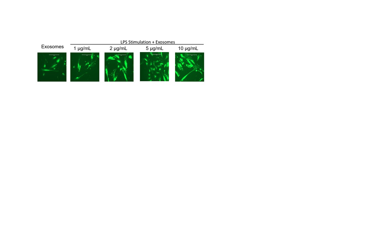 Figure 3. EVOS imaging showing the uptake of ExoGlow™-labeled blueberry-derived exosomes by normal colon CCD841 CoN cells under stimulation by varying concentrations of lipopolysaccharide (LPS). The scale bar represents 200 μm.