
Formulation and Delivery - Biomolecular
Category: Poster Abstract
Priyal Bagwe, BS (she/her/hers)
Mercer University
Atlanta, Georgia, United States
Priyal Bagwe, BS (she/her/hers)
Mercer University
Atlanta, Georgia, United States
Sarthak Shah, MS (he/him/his)
PhD Student
Mercer University
atlanta, Georgia, United States
Susu Zughaier, Ph.D. (she/her/hers)
Qatar University
Doha, Ad Dawhah, Qatar
Martin J. D'Souza, Ph.D.
Mercer University
Atlanta, Georgia, United States
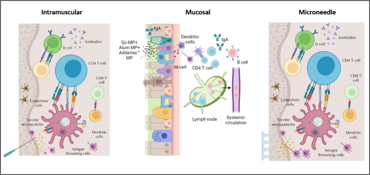 Figure 1. Exploring pain-free vaccination alternatives for a whole cell inactivated microparticulate gonorrhea vaccine. The mucosal immune system consists of specialized cells, including T and B lymphocytes, antigen-presenting cells, and secretory IgA antibodies. The primary function of mucosal immunity is to provide protection against pathogens that enter the body through mucosal surfaces. Unlike the systemic immune response, which mainly produces IgG antibodies, mucosal immunity produces IgA antibodies, which are secreted into the mucosal secretions and can neutralize pathogens before they can establish an infection. Transdermal immunization using dissolving microneedles is another attractive pain-free alternative. The skin contains specialized antigen-presenting cells, including Langerhans cells, dermal dendritic cells, and resident macrophages, making it a promising site for immunization. Langerhans cells play a crucial role in immune surveillance and signaling to T cells, which are drained into nearby lymph nodes, stimulating B cells to produce antibodies.
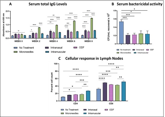 Figure 2. A. Microneedle, intranasal, buccal, and intramuscular immunization with whole-cell inactivated gonococci vaccine MP generated antibodies against N. gonorrhoeae. The mice were immunized with three doses of Gc-MP (100 µg) combined with adjuvants (Alum MP or AddaVax™ MP). Dilution Titer- 1:100. B. Serum Bactericidal Assay. Colony Forming Units recovered (CFU/mL) of N. gonorrhoeae exposed to mice sera at 1:1000 dilution. Data are expressed as mean ± SEM, N=5 mice, Two-way repeated measures ANOVA, * p < 0.05 significant, ** p < 0.01 very significant, *** p < 0.001 extremely significant, **** p < 0.0001 extremely significant. C. Expression of CD4 and CD8 on the surface of the lymph node cells from vaccinated mice. The mice were sacrificed two weeks after the challenge study's beginning. The surface expression of the CD4 Lymph node and CD8 Lymph node induced by MN, ODF, IN, and IM immunization was compared to no treatment control cells. Data are expressed as Mean ± S.E.M., N=5 mice, *** p < 0.001 extremely significant, ** p < 0.01; * p < 0.05 (Brown Forsythe ANOVA test).
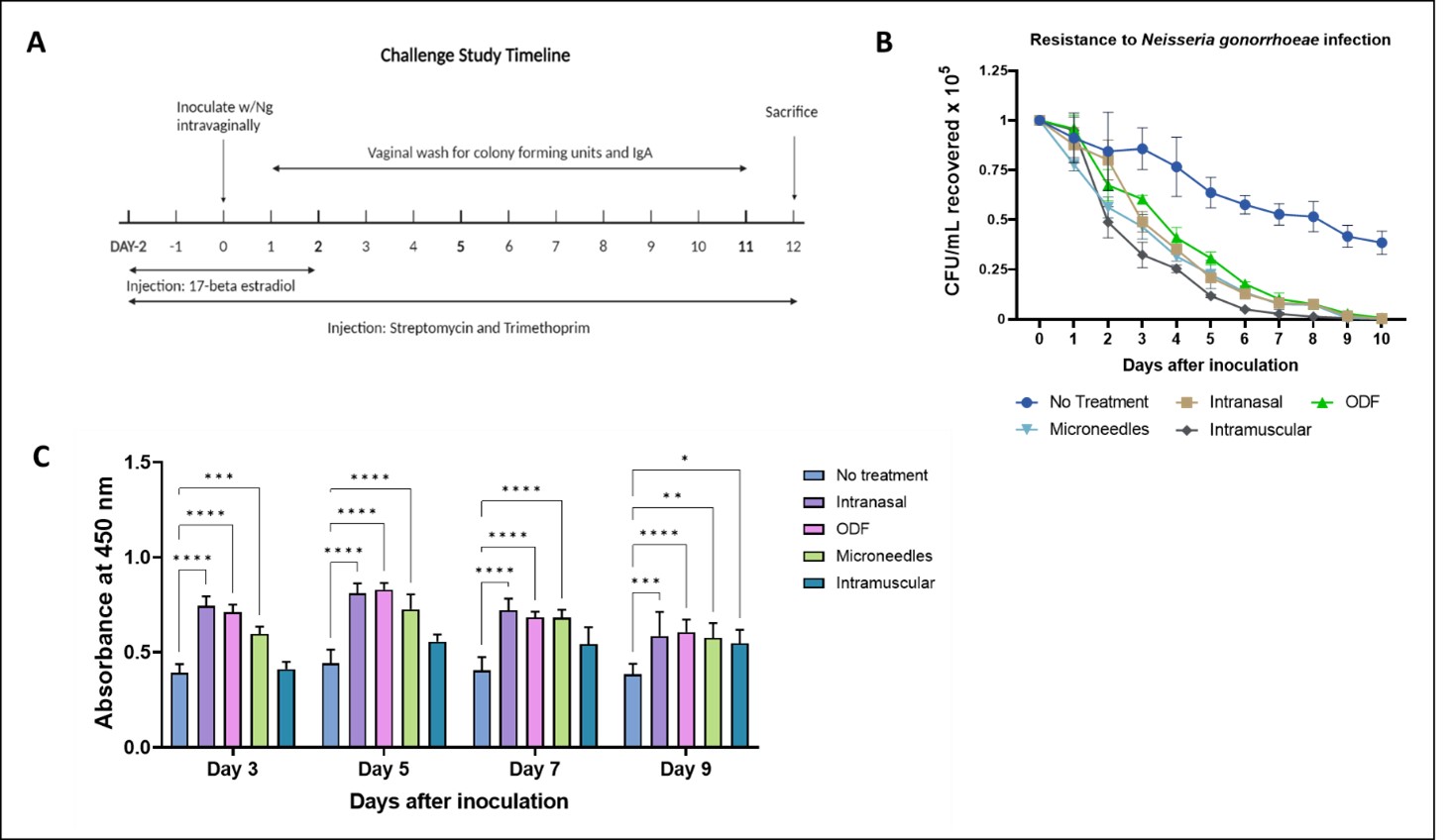 Figure 3. (A) Challenge Study Timeline: The mice were challenged at week 8 intra-vaginally with a live Neisseria gonorrhoeae strain, CDC-F62, in the murine lower genital tract infection model at 106 CFU/mL. The infection was monitored by analyzing vaginal washes cultured on agar plates. (B) Resistance to live bacterial infection in mice following challenge, CFU/mL recovered of N. gonorrhoeae from the mice immunized with the vaccine in MN, ODF, IN, and IM routes were compared to the control group. (C) Local vaginal IgA Levels (Absorbance at 450nm). Data are expressed as mean ± SEM, N=5 mice, **** p < 0.0001, *** p < 0.001, ** p < 0.01, * p < 0.05 (ANOVA, no treatment vs. IM, IN, ODF, MN).