
Discovery and Basic Research
Category: Poster Abstract
Sreelakshmi Nandakumar Menon, MS (she/her/hers)
Mercer University
Atlanta, Georgia, United States
Sreelakshmi Nandakumar Menon, MS (she/her/hers)
Mercer University
Atlanta, Georgia, United States
Morgan Daniel, Pharm.D. (she/her/hers)
Mercer University
Atlanta, Georgia, United States
Emmanuella Ezewudo, BS (she/her/hers)
Mercer University
Atlanta, Georgia, United States
Farzana Zerin, BS (she/her/hers)
Mercer University
Atlanta, Georgia, United States
Nimi Simon, BS (she/her/hers)
Mercer University
Atlanta, Georgia, United States
Nader Moniri, Ph.D. (he/him/his)
Mercer University
Atlanta, Georgia, United States
Raquibul Hasan, Ph.D. (he/him/his)
Mercer University
Atlanta, Georgia, United States
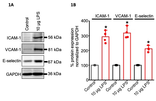 Fig. 1: LPS treatment stimulates the over expression of adhesion molecules in mouse mesenteric artery endothelial cells (MECs). (A) Western blot images illustrating induction of ICAM-1, VCAM-1 and E-selection proteins by LPS in MECs. (B) Mean data for ICAM-1, VCAM-1 and E-selection protein expression. n=4
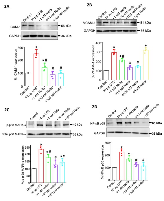 Fig. 2: (A) Neflamapimod inhibits LPS-stimulated induction of ICAM-1 on MECs. Representative Western blot images showing ICAM-1 induction by LPS and inhibition of ICAM-1 expression by 1, 10 and 100 nM Neflamapimod treatment in MECs. Mean data for ICAM-1 protein expression. n=4. *p < 0.05 vs Control; #p < 0.05 vs LPS-treated group.
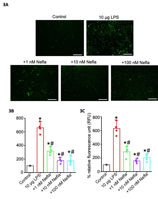 Fig. 3: Neflamapimod inhibits leukocyte (THP-1 cells) adhesion onto MECs. (A) Immunofluorescence images comparing attached THP-1 cells (green) onto MECs in LPS- and Neflamapimod -treated groups with untreated controls. (B) Mean data for the percentage attached THP-1 cells onto MEC monolayer, n=4. (C) Mean RFU of solubilized cells, n=4. * p < 0.05 vs Control, # p < 0.05 vs LPS. Scale bar in the images = 50 µm.