
Formulation and Delivery - Biomolecular
Category: Poster Abstract
Apoorva Daram (she/her/hers)
St. John's University
Fresh Meadows, New York, United States
Apoorva Daram (she/her/hers)
St. John's University
Fresh Meadows, New York, United States
Shruti Sawant (she/her/hers)
Purdue University
Queens, New York, United States
Vasudha Prithipaul, B.S. (she/her/hers)
St. John's University
Queens, New York, United States
Nitesh Kunda, Ph.D.
St. John's University
New York, New York, United States
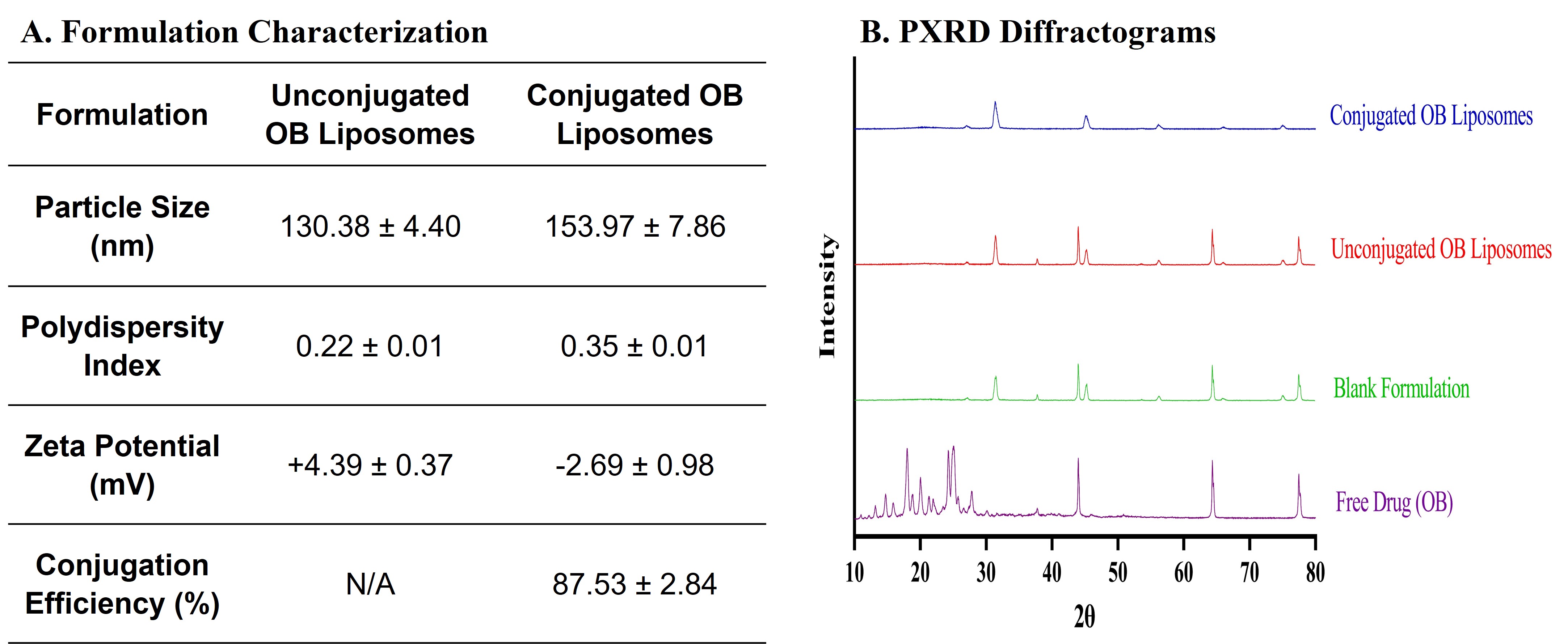 Figure 1. Immunoliposome formulation development. A. Physiochemical characterization of Osimertinib Immunoliposomes (Conjugated OB Liposomes). Data represents mean ± SD (n = 5). B. The XRD diffractogram of OB, blank formulation, unconjugated OB liposomes, and conjugated OB liposomes.
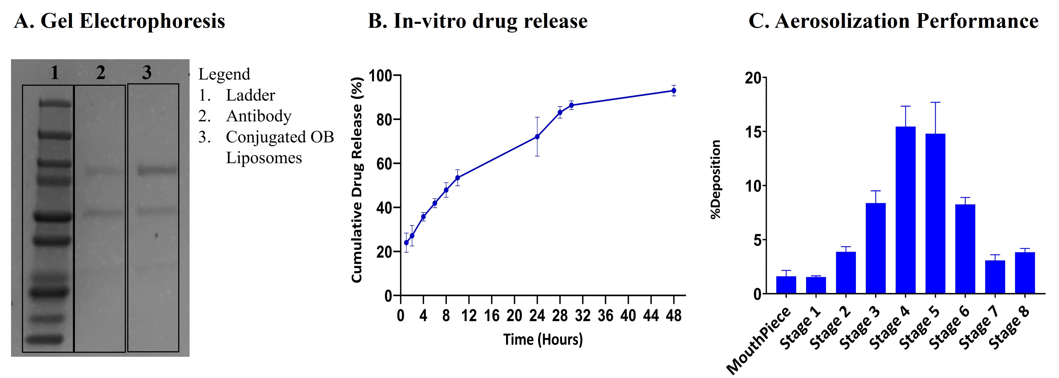 Figure 2. Immunoliposome characterization. A. Gel electrophoresis/Coomassie blue staining of free antibody and conjugated OB liposome samples. B. Cumulative release profile for OB from immunoliposomes in phosphate buffer saline (PBS), pH 7.4. Data represents mean ± SD (n = 4) C. In-vitro aerosol deposition profile represented as percent of drug deposited on each stage of Next Generation Impactor (NGI). Data represents mean ± SD (n = 3)
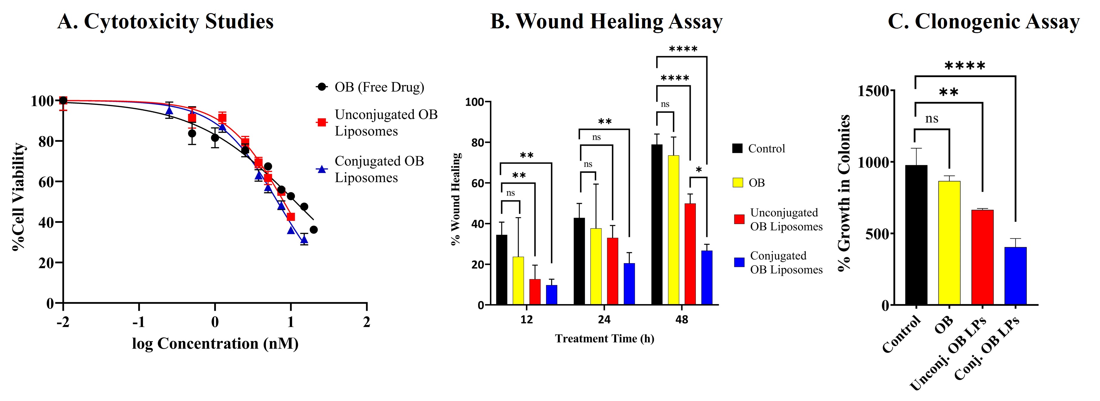 Figure 3. Cytotoxicity and Migration Assay. A. Cytotoxicity studies after 72h treatment with OB, unconjugated and conjugated OB liposomes determined using MTT assay in H1975 cell line. B. Scratch assay analysis of H1975 cell line, shown as % wound healing over time, after treatment with OB, unconjugated and conjugated OB Liposomes. C. Clonogenic assay analysis after 72h of OB, unconjugated and conjugated OB liposomes. Data represents mean ± SD (n = 3)