
Formulation and Delivery - Chemical
Category: Poster Abstract
Riddhi Trivedi, BS (she/her/hers)
Graduate student
North Dakota State University
Fargo, North Dakota, United States
Riddhi Trivedi, BS (she/her/hers)
Graduate student
North Dakota State University
Fargo, North Dakota, United States
Jagdish Singh, Ph.D. (he/him/his)
North Dakota State University
Fargo, North Dakota, United States
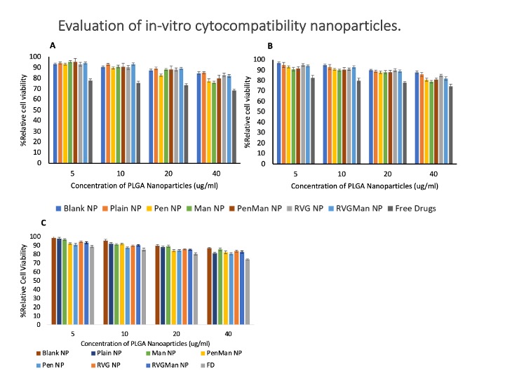 : In-vitro cytocompatibility of different PLGA nanoparticle formulations and free drugs in (A) brain endothelial cells, (B) primary astrocytes and (C) microglia cells. The cellular viability of free drug and PLGA at different concentrations was evaluated upon exposure for 24 h Data represented as mean ± S.D. (n = 6).
  Quantitative cellular uptake of different PLGA nanoparticles and free drugs in bEnd.3 cells (A) and primary astrocytes (B) at 0.5, 1, 2, 4, and 6h. Data represented as mean ± S.D. (n = 6). At 6 h, drug concentration in the cells was significantly higher than all the earlier time points when treated with PenMan NP *(p < 0.05), # PenMan NP was significantly higher compared to other formulations at 6 h. In-vitro release of Emtricitabine (A) and Bictegravir (B) from PLGA Nanoparticles. Formulations were prepared using 40% w/w and 10% w/w drug/polymer ratio, respectively. Data represented as mean ± S.D. (n = 6). No significant difference in drug release was noted between the formulations at all time points.
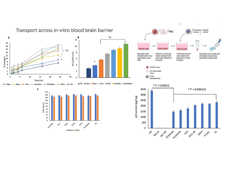 Transport through BBB model over a period of 24 h. (B) Endothelial cell permeability coefficient (Pe, expressed in 1x10⁻⁶ cm/s). (C) Measurement of TEER of BBB model before and after 24 h of transport study upon incubation with different PLGA nanoparticles formulations and free drug. Data represented as mean ± S.D. (n = 4). Statistically significant differences (p < 0.05) is shown as (*) with Free drug and (#) Plain nanoparticles. In-vitro viral suppression after treatment with different PLGA nanoparticles and free drug. Data represented as mean ± S.D. (n = 4). Statistically significant differences shown in graph.