
Formulation and Delivery - Biomolecular
Category: Poster Abstract
.jpg "Bishal Misra, MS (he/him/his) photo")
Bishal Misra, MS (he/him/his)
Graduate Student
West Virginia University
Morgantown, West Virginia, United States
Sharan Bobbala, Ph.D. (he/him/his)
West Virginia University
Morgantown, West Virginia, United States
.jpg) Fig. 1. Physico-chemical characterization of adjuvanted LNPs. A) Particle size of the LNPs measured using dynamic light scattering and number average is reported as d.nm. Negative stained TEM images of CL347-SM102 and CL347 only are reported along with it. The scale is 100 nm. B) Encapsulation efficiency of mCherry mRNA of the CL347 LNPs and CL347-SM102 LNPs measured by QuantiFluor® RNA System. Data represented as mean ± SD (n=4).
Fig. 1. Physico-chemical characterization of adjuvanted LNPs. A) Particle size of the LNPs measured using dynamic light scattering and number average is reported as d.nm. Negative stained TEM images of CL347-SM102 and CL347 only are reported along with it. The scale is 100 nm. B) Encapsulation efficiency of mCherry mRNA of the CL347 LNPs and CL347-SM102 LNPs measured by QuantiFluor® RNA System. Data represented as mean ± SD (n=4). 
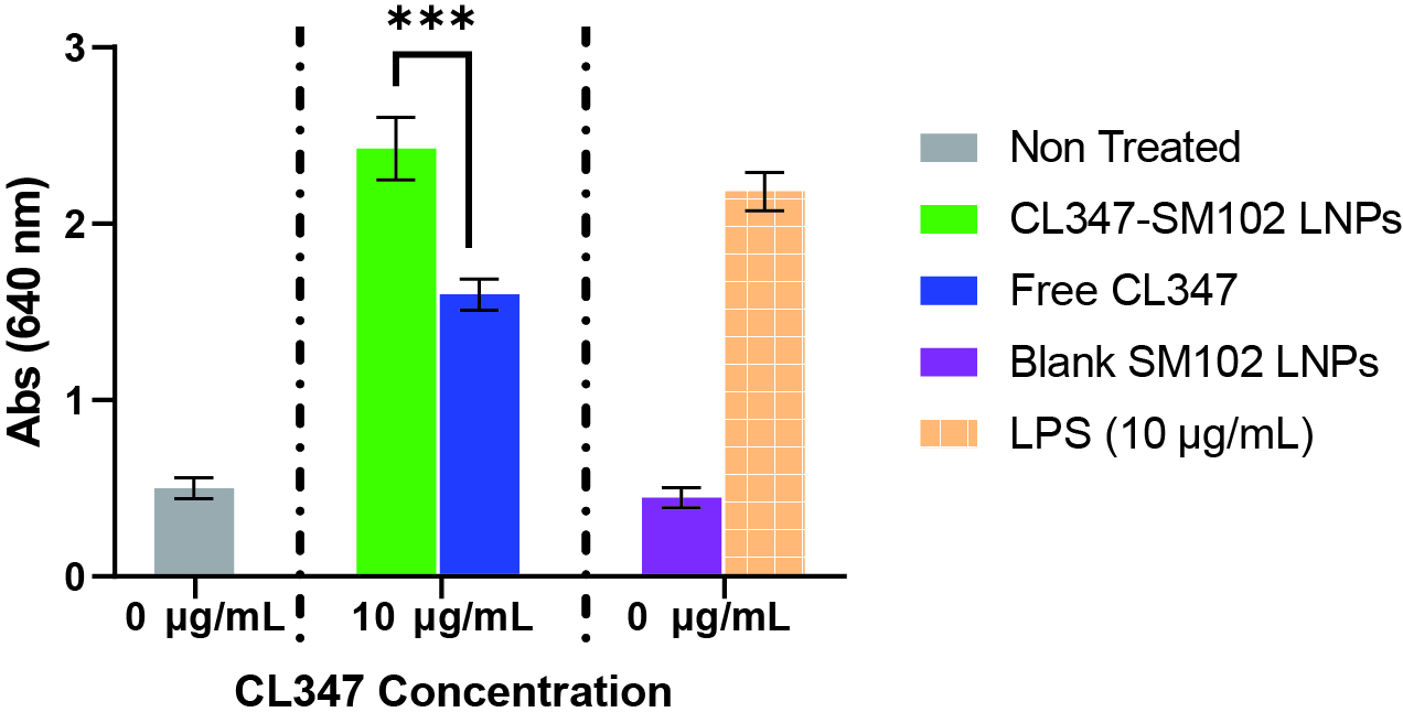 Fig. 3. Secreted embryonic alkaline phosphatase (SEAP) activity of CL347 LNPs after 48 hours of treatment with Raw-Blue™ Cells. Y-axis indicates absorption intensity at 640 nm, directly proportional to the SEAP activity of the samples containing CL347 in LNPs. Data represented as mean ± SD (n=3). Significant differences between each timepoint were determined by one way ANOVA with Tukey’s multiple comparison test, ****p < 0.0006.