
Formulation and Delivery - Chemical
Category: Poster Abstract
M. Vitoria Lopes Badra Bentley, Ph.D. (she/her/hers)
University of Sao Paulo
Ribeiro Preto, Sao Paulo, Brazil
Daniel Giuliano Giuliano Cerri, Ph.D. (he/him/his)
Universidade de Sao Paulo
Ribeirao Preto, Sao Paulo, Brazil
Fabíola Silva Garcia Garcia (she/her/hers)
Universidade de Sao Paulo
Ribeirao Preto, Sao Paulo, Brazil
Emanuel Carrilho, Ph.D. (he/him/his)
Universidade de Sao Paulo
Sao Carlos, Sao Paulo, Brazil
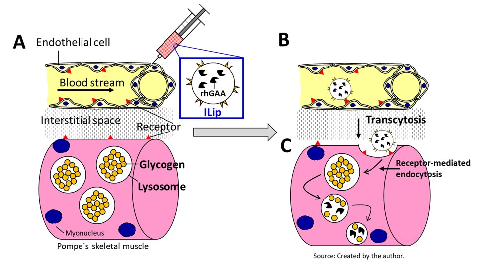 Figure 1. Immunoliposome (Ilip) should be injected intravenously (A), where it will bypass the endothelial tissue by transcytosis (B) , the interstitial space by Brownian movement and interacting with specific muscle receptor (C) by activating receptor-mediated endocytosis, which will deliver the enzyme to the lysosome.
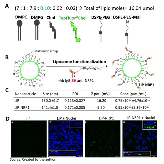 Figure 2. (A) Fluorescent liposome composition; (B) Liposome functionalization with partially reduced anti-NRP1; (C) Physicochemical data obtained from control liposome (L488) and functionalized one (L488-NRP1) and; (D) Liposomes interaction with pre-fixed differentiated myotubes via confocal fluorescence microscopy, where (a and b)= L488 and (c and d)= L488-NRP1 and; (a) and (c) liposomes excitation only and (b) and (d) liposomes merged with nuclei staining and upper and bottom insets of lateral view. Note that only L488-NRP1 (d) showed a strong interaction with cell plasma membrane surface.
 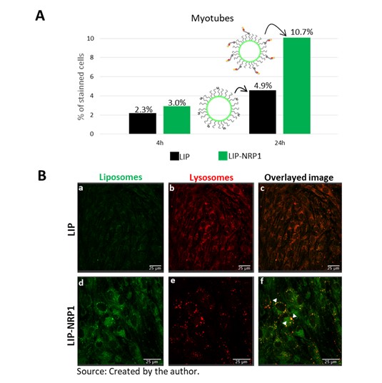 Figure 3. Liposomes dynamic in uptake assays. (A) By Flow cytometry, it was showed that LIP-NRP1 is uptake practically the double than LIP after 24h of exposition and; (B) Confocal fluorescent microscopy of 24h exposition of liposomes into myotubes, where: (a-c) LIP, (d-f) LIP-NRP1; (a and d) Liposomes staining; (b and e) lysosome staining and (c and f) overlaid images. Note that LIP-NRP1 was delivered inside myotubes lysosomes as seen by the yellow staining in the overlaid image (f, arrowheads).