
Formulation and Delivery - Biomolecular
Category: Poster Abstract
Sruthi Sarvepalli, MS (she/her/hers)
Doctoral Fellow
St. John's University
newark, New Jersey, United States
Sruthi Sarvepalli, MS (she/her/hers)
Doctoral Fellow
St. John's University
newark, New Jersey, United States
Ketan Patel, Ph.D.
St. John's University
Jamaica, New York, United States
Sandra Reznik, M.D., Ph.D.
St. John's University
Queens, New York, United States
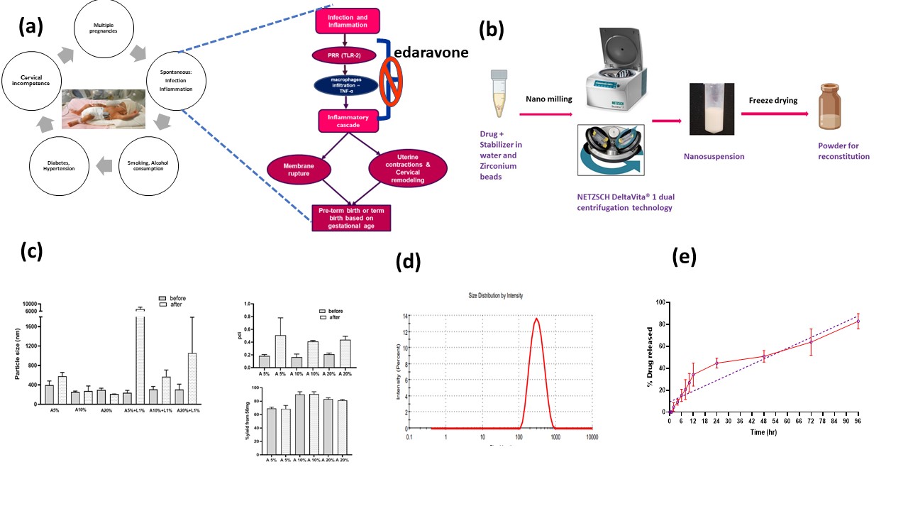 Fig 1. Preterm labor pathophysiology, Development and characterization of Edaravone Nanosuspension. (a) preterm birth causes and pathophysiology (b) Schematic presentation of formulation preparation. (c) Characterization including particle size, pdi, %nano yield of all the 6 shortlisted formulations before and after freeze drying. (d) Dynamic light scattering graph illustrating unimodal particle size distribution of the final formulation that is stabilized by BSA 10%. (e) Cumulative % in vitro release profile of final formulation demonstrating controlled release of edaravone.
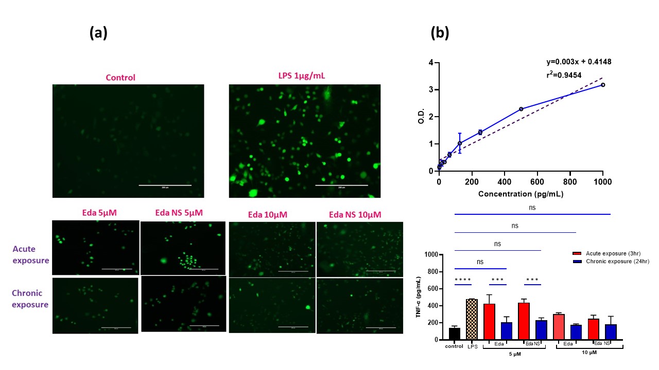 Fig 2. In vitro biological efficacy studies of the Eda NS in RAW 264.7 cells. (a) Pictures representing reduction in LPS induced ROS associated fluorescence with treatments upon ROS sensitive staining [by Chloromethyl derivative of dihydro dichloro fluorescein diacetate, acetyl ester (CM-H₂DCFDA)] of the cells. (b) TNF-α linearity, reduction in LPS induced TNF-α levels, [significant with chronic exposure than acute (at 5µM, p < 0.001)] with treatments - ELISA study.
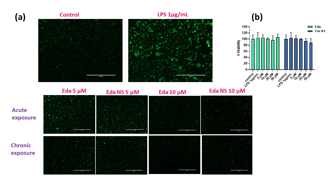 Fig 3. In vitro biological efficacy studies of the Eda NS in HTR-8/SVneo cells. (a) Pictures representing reduction in LPS induced ROS associated fluorescence with treatments upon ROS sensitive staining (CM-H₂DCFDA) of the cells. (b) Safety profile of the pure drug and Eda NS with >85% cell viability is proven by MTT study.