
Manufacturing and Analytical Characterization - Biomolecular
Category: Poster Abstract
Irene Chang, Ph.D. (she/her/hers)
Merck & Co., Inc.
Lansdale, Pennsylvania, United States
Irene Chang, Ph.D. (she/her/hers)
Merck & Co., Inc.
Lansdale, Pennsylvania, United States
Michael Winters, Ph.D.
Merck & Co., Inc.
West Point, Pennsylvania, United States
Christopher Farrell, Ph.D.
Merck & Co., Inc.
West Point, Pennsylvania, United States
Michael McNevin, Ph.D.
Merck & Co., Inc.
West Point, Pennsylvania, United States
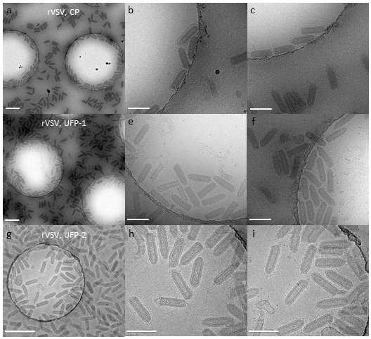 Figure 1. CryoTEM of rVSV process intermediates CP (a to c), UFP-1 (d to f), and UFP-2 (g to i). See the text for sample detail. All three samples consisted of mostly bullet-shaped particles, but CP and UFP-2 contained fewer nucleocapsids without a surrounding membrane or had undergone uncoiling, as well as a denser layer of surface spike proteins, than UFP-1. Scale bars: 500 nm in a, d, and g; 200 nm in rest of the figures.
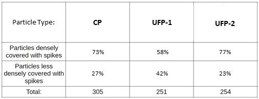 Table 1. Fraction counting analysis of bullet-shaped particles densely covered with spike proteins in rVSV process intermediates. See the text for sample detail. Particles categorized as densely covered (multiple stretches of membrane 50 nm long containing ≥3 S proteins) or less densely covered with the protein. UFP-2, generated by concentration of CP material using a low shear pump, contained a higher percentage of particles densely covered with spikes compared to UFP-1, generated using a default pump. UFP-2 and CP had comparable percentages of particles densely covered with spikes.
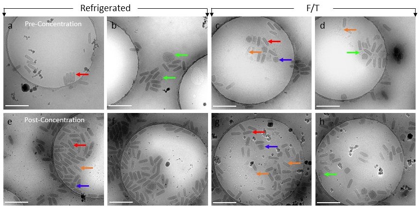 Figure 2. CryoTEM images of a rVSV vaccine material subjected to refrigeration (refrigerated) or a freeze-thaw (F/T) cycle. The material was imaged neat (pre-concentration, a to d) or after concentration by ultracentrifugation (post-concentration, e to h). See the text for sample detail. After concentration, the refrigerated material had mostly bullet-shaped particles (red arrow) typical of VSV, but also a few round particles (blue arrow). The F/T material consisted of a mixed population of bullet-shaped and round particles even before sample concentration. Empty vesicles (green arrow) and un-enveloped and unwound nucleocapsid (orange arrow) were also present in some of the samples. Scale bars: 500 nm.