
Formulation and Delivery - Chemical
Category: Poster Abstract
Iria Seoane-Viano, Ph.D.
University College London
London, England, United Kingdom
Iria Seoane-Viano, Ph.D.
University College London
London, England, United Kingdom
Jun Jie Ong, B.S. (he/him/his)
University College London
LONDON, England, United Kingdom
Tania Pérez-Ramos, M.D.
University Hospital Lucus Augusti
LUGO, Galicia, Spain
Elena Guerra-Baamonde, M.D.
University Hospital Lucus Augusti
LUGO, Galicia, Spain
Jiaqi Liu, M.S. (she/her/hers)
PhD Researcher
University College London
LONDON, England, United Kingdom
Jorge Gonzalez-Ramirez, M.D.
University Hospital Lucus Augusti
LUGO, Galicia, Spain
Manuel Vazquez-Caruncho, M.D.
University Hospital Lucus Augusti
LUGO, Galicia, Spain
Alvaro Goyanes, Ph.D.
FabRx Ltd.
London, England, United Kingdom
Abdul Basit, Ph.D.
University College London
London, England, United Kingdom
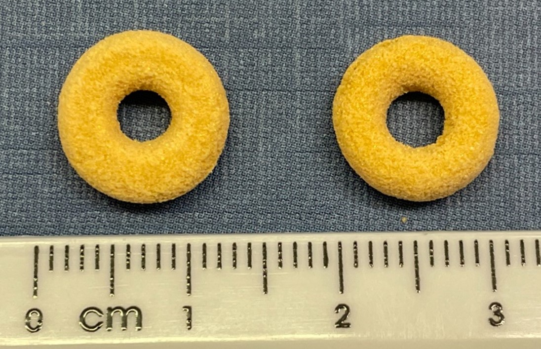 Figure 1. On the left SLS90 and on the right SLS130.
.jpg) Table 1. In vivo and in vivo disintegration times.
Table 1. In vivo and in vivo disintegration times.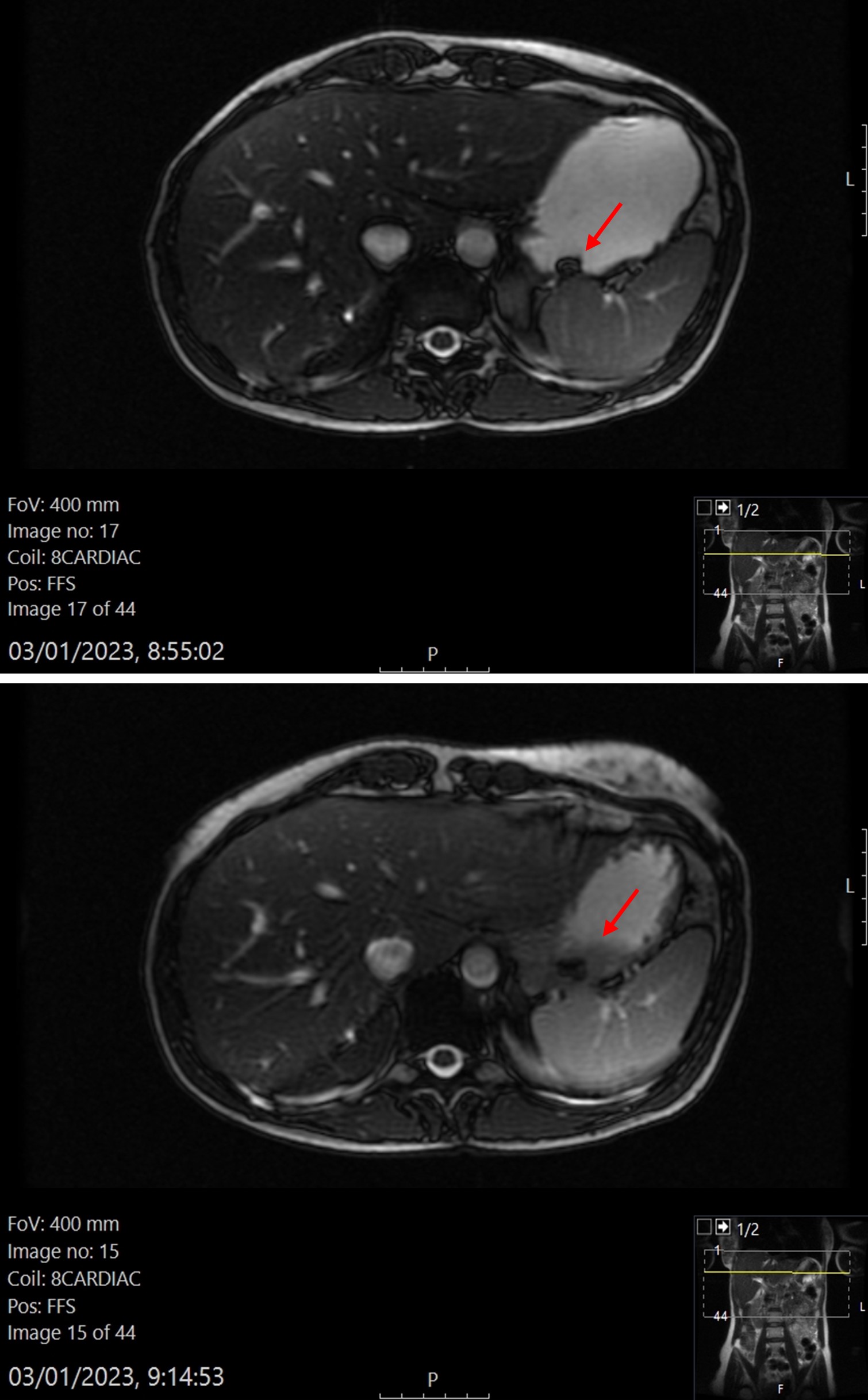 Figure 2. Representative MRI images of the SLS90 formulation. At the top, image taken after ingestion of the formulation. The torus shape of the printlet can be appreciated. At bottom, image taken after the disintegration of the printlet began. A black trail as a result of tablet disintegration can be seen on the image.