
Formulation and Delivery - Biomolecular
Category: Poster Abstract
 photo")
Aishwarya Saraswat, PhD (she/her/hers)
Senior Scientist
Regeneron Pharmaceuticals, Inc.
Tarrytown, New York, United States
Aishwarya Saraswat, Ph.D.
St. John's University
jamaica, New York, United States
Hari Priya Vemana, Ph.D. (she/her/hers)
St. John's University
JAMAICA, New York, United States
Ketan Patel, Ph.D.
St. John's University
Jamaica, New York, United States
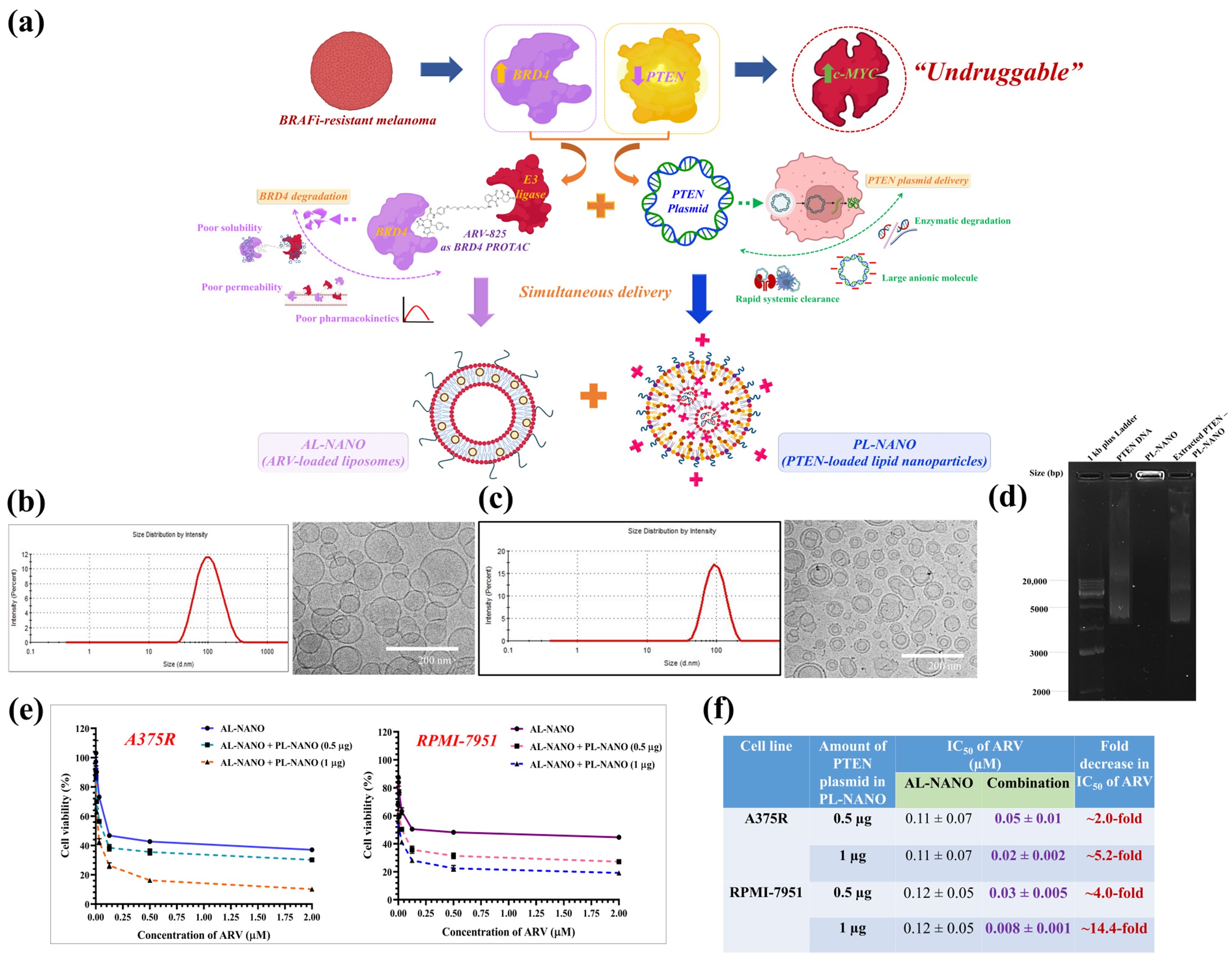 Fig. 1. (a) Graphical representation of a novel gene therapy approach for BRAFi-resistant melanoma targeting downregulation of BRD4 oncoprotein via ARV-loaded liposomes (AL-NANO) and upregulation of PTEN tumor suppressor via PTEN plasmid-loaded lipid nanoparticles (PL-NANO) for their simultaneous delivery. (b) Dynamic light scattering graph and cryo-TEM image illustrating unimodal particle size distribution of AL-NANO. (c) Dynamic light scattering graph and cryo-TEM image illustrating unimodal particle size distribution of PL-NANO and representative agarose gel electrophoresis image illustrating binding efficiency and complete entrapment of PTEN plasmid within PL-NANO. (d) MTT cytotoxicity curves in A375R and RPMI-7951 cell lines indicating strong synergism between AL-NANO and PL-NANO in acquired and intrinsically BRAFi-resistant melanoma cell lines. (e) IC50 values of ARV in AL-NANO alone and in combination with PL-NANO in BRAFi-resistant melanoma cell lines illustrating synergistic anticancer activity.
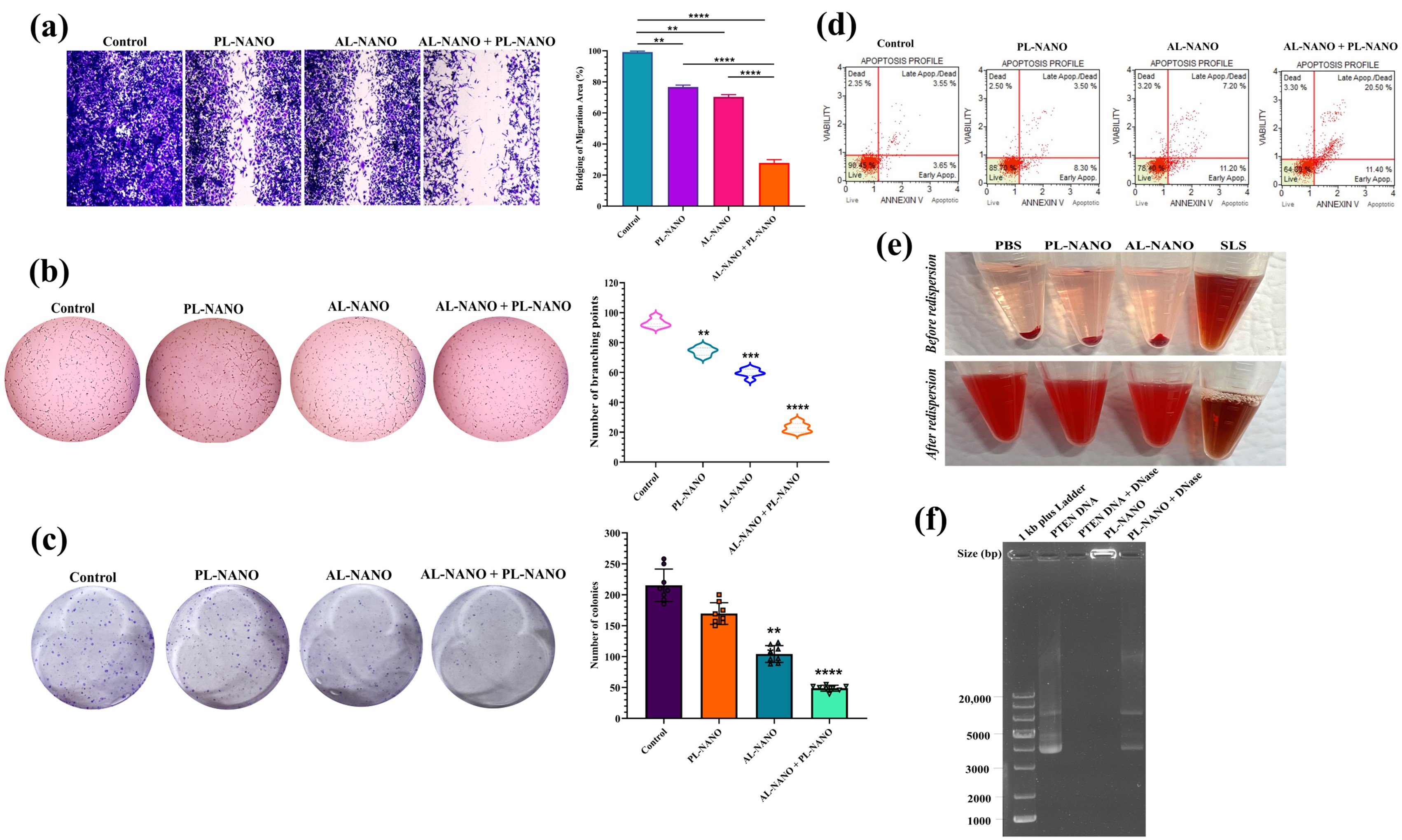 Fig. 2. (a) Microscopic images of scratch assay and percentage inhibition of migration produced by PL-NANO, AL-NANO, and PL-NANO + AL-NANO as compared to control in A375R cells. (b) Vasculogenic mimicry images and quantitative analysis of number of branching points formed in each treatment group as compared to control in A375R cells. (c) Representative images and quantitative illustration of percentage decrease in the number of colonies formed with individual and combination treatment as compared to control in A375R cells. (d) Flow cytometry analysis results depicting significant apoptotic effect of PL-NANO and AL-NANO combination resulting in 31.9% apoptotic cell population in A375R cells. (e) Negligible hemolysis demonstrated by PL-NANO and AL-NANO as tested in mice RBCs. (f) DNase protection assay results illustrating prevention of PTEN-DNA from enzymatic degradation by PL-NANO using agarose gel electrophoresis. **p < 0.01, ***p < 0.001, ****p < 0.0001.
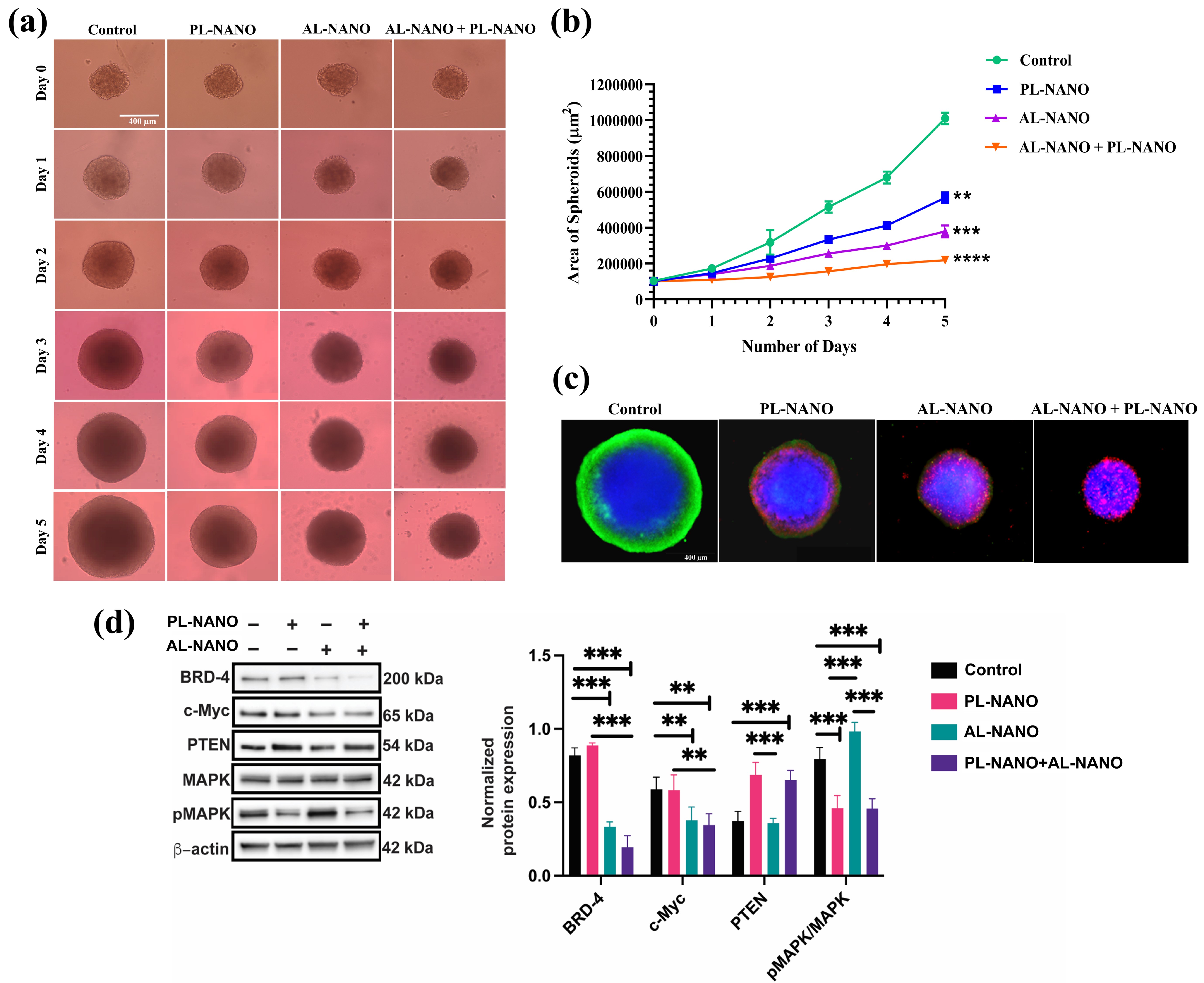 Fig. 3. (a) Representative bright field images of A375R 3D spheroids treated with PL-NANO, AL-NANO, and PL-NANO + AL-NANO during 5 days of treatment. (b) Comparison of the area of spheroids treated with different groups during 5 days of treatment. Significant difference was observed in the area of spheroids treated with PL-NANO, AL-NANO and PL-NANO + AL-NANO in comparison to control. (c) Fluorescence images signifying apoptosis of spheroids treated with various groups. Composite images of DAPI (blue), Calcein AM (green) and Ethidium Homodimer-1 (red). Scale bar: 400 μm. (d) Representative western blots for expression of BRD4, c-Myc, PTEN, MAPK and pMAPK proteins and quantification of the western blot results for expression of each target protein in A375R cells. **p < 0.01, ***p < 0.001, ****p < 0.0001.